Structural Analysis and
Modeling Tools for Cryo-EM Density Maps at Medium Resolutions
 PI:
Jing He
PI:
Jing He
Co-PI: Desh Ranjan, Mohammed Zubair, Shuiwang Ji
Department of Computer Science
Old Dominion University
Web page: http://www.cs.odu.edu/~jhe
Funding support: NSF DBI-1356621
Secondary Structure Tracing from Cryo-EM
Density Maps at Medium Resolutions
Cryo-electron Microscopy (Cryo-EM) is a biophysical technique to derive 3-dimensional density maps at various resolutions. Atomic structures can be obtained from high resolution density maps such as those beyond 3.5 angstroms. However, when density maps are resolved at medium resolutions, such as those 4-8 angstrom, it is still challenging to derive atomic structures. The most visible structural components in a medium-resolution density map are secondary structures such as alpha-helices and beta-sheets. Since secondary structures are the major components of a protein structure, automatic detection of secondary structure from a density map is an important step in structural analysis.
Tool Download - SSETracer
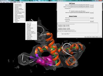
SSETracer is a tool to detect secondary structure locations from a 3-demensional density map at medium resolutions. Currently it is integrated in Chimera.
Tracing Beta Strands from a Beta-sheet Image
Although it is possible to detect beta-sheets from a cryo-EM density map at medium resolutions, it is a challenging problem to detect beta-strands due to close spacing of beta-strands. Two neighboring beta-strands are 4.5-5 angstroms apart, and therefore they are often not resolved in a density map with a lower than 4.5 angstrom resolution. However, it is possible to predict the traces of beta-strands through modeling the twist, a unique character of beta-strands.
Tool Download - StrandTwister
StrandTwister is a tool to predict traces of beta-strands from an isolated beta-sheet density image. Currently it is integrated in Chimera together with SSETracer.
Exit Questionnaire for Past Research Experience - Exit Questionnaire